Clinical Expertise
Dr. Sylvia Shitsama brings more than two decades of specialist medical experience to consultations and surgical interventions across five primary clinical disciplines.
Brain Trauma
Comprehensive clinical evaluation and urgent neurosurgical intervention for acute head injuries, traumatic brain injury (TBI), cranial fractures, and intracranial bleeding resulting from road traffic accidents, falls, or occupational injuries.
Clinical & Surgical Scope
- Acute Epidural & Subdural Hematoma Evacuation
- Decompressive Craniectomy for Refractory Intracranial Pressure
- Depressed & Comminuted Skull Fracture Elevation
- Cranioplasty & Post-Traumatic Cranial Reconstruction
- Post-Concussion Assessment & Monitoring
Patient Guidance & Common Queries
Q:When is surgical intervention required following a head injury?
Surgery is indicated when urgent CT neuroimaging demonstrates significant intracranial hemorrhage (such as epidural or acute subdural hematoma) exerting mass effect on the brain, or severely depressed skull fractures.
Q:What initial investigations are essential?
A non-contrast brain CT scan is the urgent gold-standard examination to rule out bleeding, fractures, or midline shift.

Emergency neurotrauma triage and surgical intervention protocol.
Brain Tumors
Specialist microsurgical management of benign and malignant brain tumours. Clinical practice prioritizes maximal safe tumour resection while diligently preserving neurological function and speech/motor pathways.
Clinical & Surgical Scope
- Gliomas, Astrocytomas & Glioblastoma Multiforme (GBM)
- Meningiomas (Convexity, Parasagittal & Skull Base)
- Pituitary Adenomas & Sellar Lesions
- Metastatic Brain Lesions
- Intraventricular Tumours & Colloid Cysts
Patient Guidance & Common Queries
Q:What symptoms suggest a brain tumor evaluation is needed?
Persistent new-onset headaches (especially waking headaches accompanied by morning nausea/vomiting), new seizures in adults, progressive focal weakness, vision changes, or cognitive/personality changes.
Q:What neuroimaging is required prior to consultation?
A contrast-enhanced Brain MRI (Magnetic Resonance Imaging) is typically the most definitive diagnostic scan for detailed tumour characterisation and surgical planning.
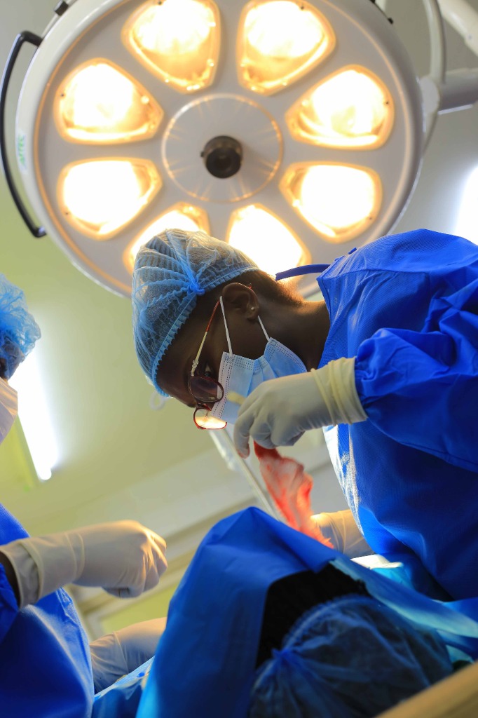
Microsurgical cranial tumour resection under high-intensity surgical illumination.
Spine
Evaluation and surgical care for conditions affecting the cervical, thoracic, and lumbar spine. Treatment follows an evidence-based pathway, ensuring conservative management is exhausted before considering surgical decompression or stabilization.
Clinical & Surgical Scope
- Cervical & Lumbar Disc Herniation (Sciatica / Radiculopathy)
- Lumbar Spinal Stenosis & Neurogenic Claudication
- Degenerative Spondylolisthesis & Vertebral Instability
- Spinal Cord Decompression & Microdiscectomy
- Spinal Trauma, Fractures & Instrument-Assisted Fusion
Patient Guidance & Common Queries
Q:When should someone consider spine surgery?
Surgery is typically considered when severe nerve compression causes progressive weakness (e.g., foot drop), intractable pain unresponsive to conservative medical therapy, or signs of spinal cord compression (myelopathy / cauda equina syndrome).
Q:What scan is best for assessing back or neck pain radiating to the legs or arms?
An MRI of the affected spinal segment (Cervical, Thoracic, or Lumbo-Sacral) provides the highest resolution imaging of spinal discs, nerve roots, and the spinal cord.
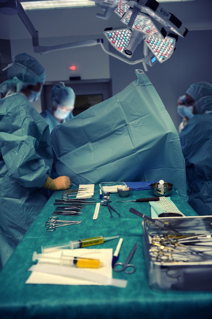
Advanced spinal decompression and stabilization within a dedicated sterile theatre environment.
Congenital Conditions
Specialist surgical repair and multidisciplinary coordination for congenital malformations of the nervous system, addressing structural anomalies from the neonatal period through childhood and adult life.
Clinical & Surgical Scope
- Spina Bifida (Myelomeningocele & Meningocele Closure)
- Encephaloceles (Occipital & Sincipital)
- Tethered Cord Syndrome & Surgical Release
- Chiari Malformations (Type I Decompression)
- Dermal Sinus Tracts & Spinal Dysraphism
Patient Guidance & Common Queries
Q:Why is early closure of myelomeningocele critical in neonates?
Prompt surgical repair within the first 24 to 48 hours after birth protects exposed neural tissue from infection (meningitis) and preserves existing neurological function.
Q:What are the signs of tethered cord syndrome?
Signs may include cutaneous markers along the spine (such as dimples, tufts of hair, or lipomas), progressive foot deformities, asymmetrical lower limb weakness, or bladder dysfunction.
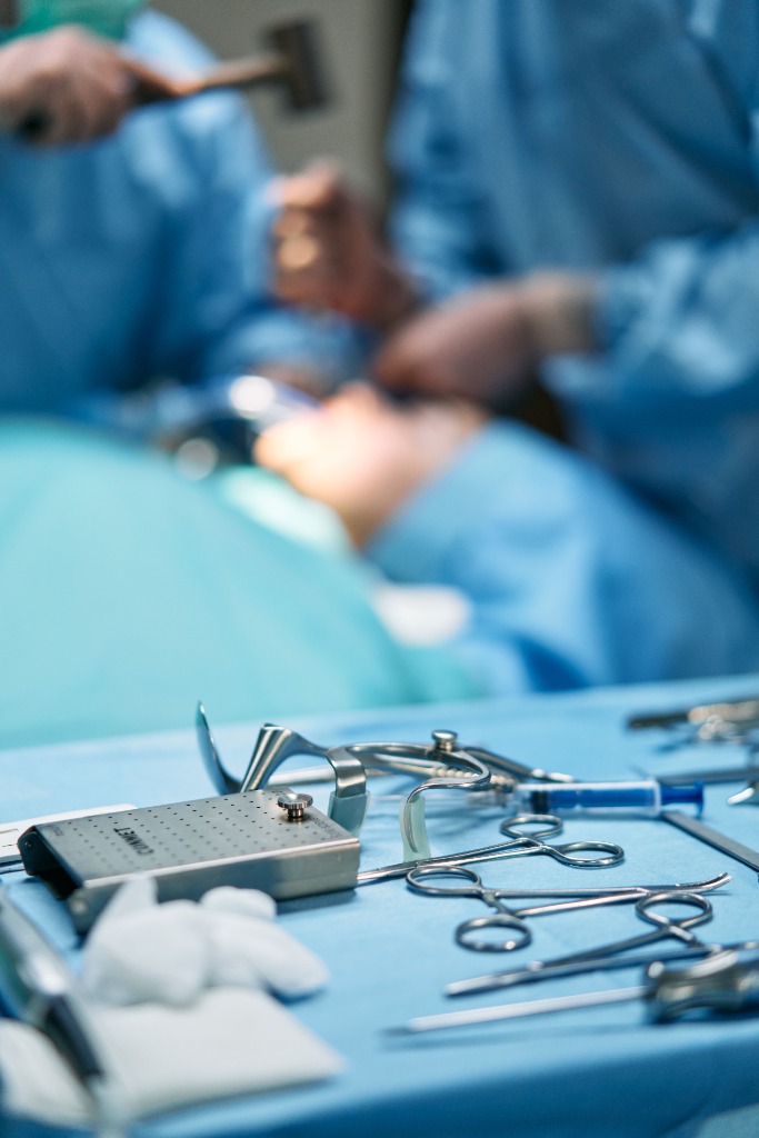
Delicate neural reconstruction using specialized micro-dissection instrumentation.
Pediatric Neurosurgery & Hydrocephalus
Gentle, compassionate, and highly specialized neurosurgical care tailored specifically to infants, children, and adolescents. This encompasses comprehensive management of pediatric hydrocephalus (ETV & VP shunts), infant head shape assessment, craniosynostosis, and childhood cranial/spinal conditions in close coordination with families.
Clinical & Surgical Scope
- Infant & Pediatric Hydrocephalus (VP Shunt Insertion & Revision)
- Endoscopic Third Ventriculostomy (ETV & ETV/CPC)
- Infant Head Shape Abnormality Assessment & Craniofacial Evaluation
- Craniosynostosis & Cranial Vault Reconstruction
- Congenital Aqueductal Stenosis & Ventricular Cysts
- Pediatric Cranial & Spinal Tumours
- Normal Pressure Hydrocephalus (NPH) & CSF Diversion
- Pediatric Neurotrauma & Accidental Fall Injuries
Patient Guidance & Common Queries
Q:What is hydrocephalus, and what signs should parents look for in infants?
Hydrocephalus occurs when excess cerebrospinal fluid (CSF) builds up within the brain’s ventricles. In infants, warning signs include an unusually rapid increase in head circumference, a bulging or tense fontanelle (soft spot), prominent scalp veins, downward-directed eyes (“sunset sign”), poor feeding, or persistent irritability.
Q:What is the difference between a VP Shunt and an ETV procedure?
A Ventriculoperitoneal (VP) Shunt diverts excess fluid from the brain to the abdominal cavity via a fine catheter and pressure-regulating valve. Endoscopic Third Ventriculostomy (ETV) is a minimally invasive neuro-endoscopic procedure that creates an internal bypass opening in the third ventricle floor, resolving blockages without a permanent shunt implant when anatomy is favorable.
Q:How is a pediatric neurosurgical consultation conducted?
Consultations are conducted in an unhurried, reassuring environment where Dr. Shitsama evaluates the child, thoroughly reviews all imaging, and answers parents’ questions in clear, accessible language.
Q:Can parents stay with their child during clinical assessments?
Yes, parents are encouraged to be present at every stage of the consultation to ensure comfort and clarity for the entire family.

Child-friendly consultation environment supporting young patients, parents, and families.
Advanced Neurosurgical Facilities & Technology
Operating in accredited tertiary neurosurgical centres equipped with precision optical magnification, continuous intraoperative neuro-monitoring, and dedicated surgical support teams.
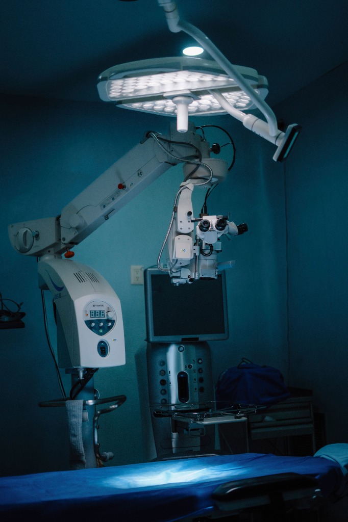
High-Resolution Neuro-Microscopy
Provides stereoscopic depth perception and intense illumination necessary for delicate microneurosurgery around critical neurovascular bundles and cranial nerves.
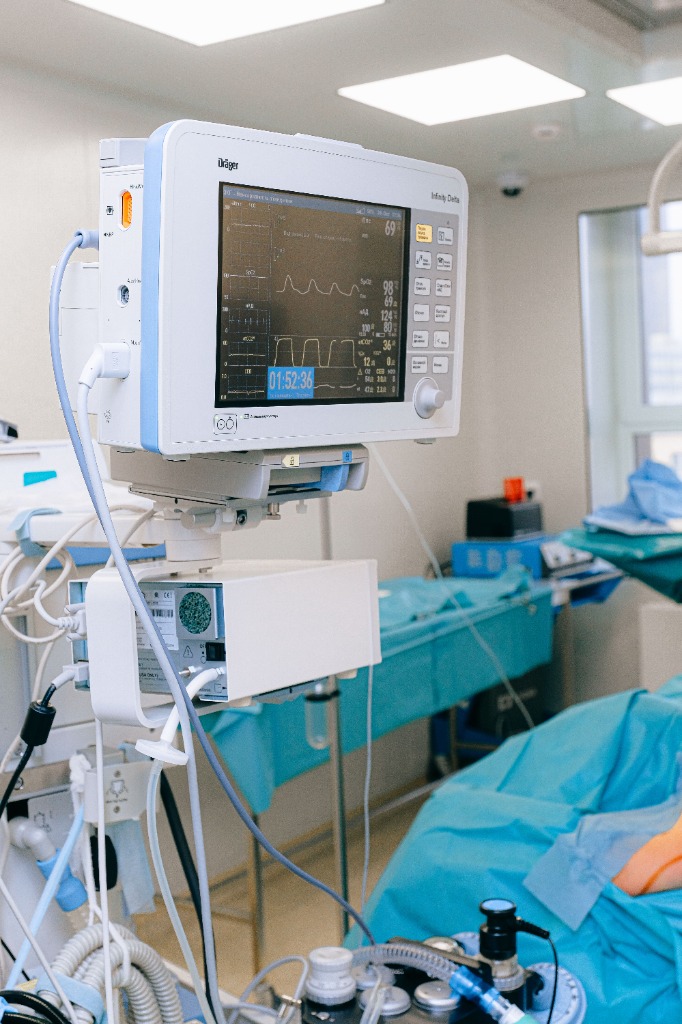
Continuous Neuro-Monitoring
Real-time physiological tracking, hemodynamic stabilization, and specialized neuro-anesthesia care safeguarding brain perfusion throughout every phase of surgery.
Sterile Field & Synchronized Team
Stringent international sterility protocols, dedicated neurosurgical scrub nursing, and meticulous preparation of microsurgical instrument sets.
Discuss Your Care with Dr. Sylvia Shitsama
Consultations take place at KNH Doctors Plaza, 4th Floor Suite 43, Hospital Road, Nairobi (Adjacent to National Bank). Inpatient surgical procedures are scheduled at affiliated accredited tertiary hospitals.